How is the procedure performed
How is the procedure performed Image-guided, minimally invasive procedures such as inferior vena cava filter placement and removal are most often performed by a specially trained interventional radiologist in an interventional radiology suite or occasionally in the operating room.
This procedure is often done on an outpatient basis. However, some patients may require admission following the procedure.
Please consult with your physician as to whether or not you will be admitted. You will be positioned on your back.
You may be connected to monitors that track your heart rate, blood pressure and pulse during the procedure.
A nurse or technologist will insert an intravenous (IV) line into a vein in your hand or arm so that sedative medication can be given intravenously.
Moderate sedation may be used. As an alternative, you may receive general anesthesia.
The area of your body where the catheter is to be inserted will be sterilized and covered with a surgical drape.
Your physician will numb the area with a local anesthetic.
A very small skin incision is made at the site.
Using image-guidance, a catheter (a long, thin, hollow plastic tube) is inserted through the skin to the treatment site.
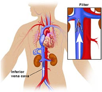
Contrast material may be injected into the inferior vena cava to help guide the catheter and verify precise placement of the IVC filter in the blood vessel.
At the end of the procedure, the catheter will be removed and pressure will be applied to stop any bleeding. The opening in the skin is then covered with a dressing. No sutures are needed.
Your intravenous line will be removed.
The procedure is usually completed within one hour.