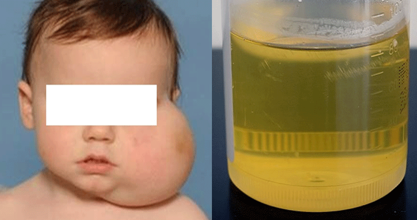
Lymphatic malformations are again within the spectrum of vascular anomalies. They are often large fluid filled spaces containing lymph as opposed to blood like venous malformations. The other type of lymphatic malformation is ‘microcystic’ and this type contains multiple tiny spaces.
The macrocystic malformations are commonly diagnosed in childhood and can grow to be very large. They can occasionally compress adjacent structures and if these are deemed important structure like for example the trachea (windpipe), treatment may be required soon.
They are treated in a similar fashion to venous malformations (ie: sclerotherapy) and similar risks are present. If the malformation is near the trachea then often a short hospital stay is required as the swelling induced by sclerotherapy can compress the trachea. This risk of this is minimal in most cases but this is discussed with the patient (or parents) prior to the procedure.
Microcystic lymphatic malformations can be more challenging to treat. There is a drug called bleomycin which has been used successfully to treat these malformations. This is a well established chemotherapy agent which is used to treat certain types of cancer (although lymphatic malformations are not cancer) and its use in much smaller doses can improve the appearance and symptoms of microcystic lymphatic malformations with excellent results. It can also be used in the treatment of other vascular anomalies.

Lymphatic malformations are associated with overgrowth (hypertrophy) and swelling of any affected area including the lips, tongue, jaws, cheeks, arms, legs, fingers or toes. Malformations affecting the tongue, windpipe (trachea) or mouth can cause difficulty breathing (dyspnea), difficulties with speech and difficulty swallowing (dysphagia) and feeding problems. If the eye socket (orbit) is involved, double vision (diplopia) or displacement of the eyeball can occur (proptosis). Lymphatic malformations affecting the chest can cause wheezing, chest pain, chest pressure, shortness of breath, difficulty breathing and potentially airway compromise. Lymphatic malformations affecting the gastrointestinal tract or pelvis can cause constipation, bladder obstruction, recurrent infection or protein loss. Lesions in bone can be associated with bone overgrowth or bone loss.
During pregnancy, a fetal ultrasound may detect some large lymphatic malformations. Ultrasound is a diagnostic tool used to evaluate organs and structures inside the body with high-frequency sound waves. After birth, diagnosis of a lymphatic malformation is generally determined by a physical examination. In addition to a complete medical history and physical examination.
Diagnostic procedures for a lymphatic malformation may include the following:
- Transillumination. A method of examination by the passage of light through tissues to assist in diagnosis. The light transmission changes with different tissues.
- Ultrasound
- CT Scan
- MRI scan
Lymphatic Malformation Treatment and Procedure Options
Lymphatic malformations are now most often treated using interventional radiology minimally-invasive (percutaneous) techniques designed to ablate (or kill) the cysts within the body. In this situation, a chemical injected into the lymphatic malformation cyst kills the cyst. The body then cleans up the dead cyst tissue over time. This can be done without large scars or the need for hospitalization.
In the past, surgical removal has been considered standard treatment for these cavernous lesions, despite significant recurrences. Percutaneous (interventional radiology) treatments for both macrocystic and microcystic lymphatic malformation now offer patients a predictable plan for both initial definitive treatment, as well as injection treatment of new cysts that fill up over time from the “solid” membranes that may be present. Microcystic injection therapy now offers patients a long-term option for percutaneous, minimally invasive treatment of new lymphatic malformation microcysts that are detected before they become problematic or develop into painful macrocysts.
Treatment options for lymphatic malformation include:
- Surgical removal
- Interventional radiology minimally-invasive ablation. Sclerotherapy.
- Rarely medical therapy (drugs: sirulimus, sildenafil) can be used to suppress the lymphatic malformation (not curative), while sclerotherapy procedures are also performed.
Bleomycin is widely used as a chemotherapy agent to treat certain types of cancer. Vascular anomalies are not cancers but bleomycin has been shown to be effective in treating certain types of vascular anomalies when injected directly into the lesion, rather than into the bloodstream as with chemotherapy. Subtypes of vascular anomalies successfully treated include, lymphatic malformations (macro and microcystic) and even venous malformations. Bleomycin works by exerting its effect on the lining of the malformation and preventing further growth and promoting regression. It does this by inhibiting local DNA synthesis. Results are encouraging with its use. This form of treatment is not widely available and often requires a course of injections over a period of months. Bleomycin treatment is considered in a multi-disciplinary setting following discussion with the patient and review of cross-sectional imaging. It can be considered as a first line treatment in micro-cystic lymphatic disease.
The risks are similar to conventional sclerotherapy in that pain and swelling are the commonest post-procedural occurrences. Occasionally ulceration can occur, especially when the lesion is close to the skin (or lining of mouth). Flu-like symptoms have also been described and usually only last for a day or so. When bleomycin is used in much higher doses and intravenously to treat cancers there is a small risk of causing lung damage. Specifically lung fibrosis has been described but when used intra-lesionally in the setting of vascular anomalies this risk is extremely small and there are no reported cases in the world. With this in mind a full assessment of lung function is carried out prior to and during a course of bleomycin treatment. If there is any suggestion of pre-existing lung problems bleomycin will not be administered.
The risks are similar to conventional sclerotherapy in that pain and swelling are the commonest post-procedural occurrences. Occasionally ulceration can occur, especially when the lesion is close to the skin (or lining of mouth). Flu-like symptoms have also been described and usually only last for a day or so. When bleomycin is used in much higher doses and intravenously to treat cancers there is a small risk of causing lung damage. Specifically lung fibrosis has been described but when used intra-lesionally in the setting of vascular anomalies this risk is extremely small and there are no reported cases in the world. With this in mind a full assessment of lung function is carried out prior to and during a course of bleomycin treatment. If there is any suggestion of pre-existing lung problems bleomycin will not be administered.
